Contact the department
- At the clinic: 0153 655311 or clinic switchboard 0153 655365
- 43 rue Cortambert – 75016 Paris – France: +33 (0)1 47 04 44 20
- Email: secretariat@imagerie-medicale-vinci.fr
- Directly via the Cortambert Centre’s website
The Victor Hugo Clinic has been home to the Leonardo da Vinci Medical Imaging Centre, located at 43 rue Cortambert in the 16th arrondissement of Paris, since 1 February 2018.
Based in the consultation building, the imaging department is equipped with an X-ray flat panel sensor table and an ultrasound scanner that is specifically suitable for the clinic’s surgical activity.
The Cortambert imaging centre is equipped with a CT scanner with X-ray dose reduction software, an MRI scanner, 4 Radiology rooms equipped for procedures, an Osteodensitometer, a dental panoramic X-ray machine, 3 Ultrasound rooms for procedures, an EOS System allowing the spine and limbs to be examined at lower doses, and a digital mammography machine coupled with an ultrasound scanner. This mammography machine is also equipped with a macrobiopsy device.
The Léonard da Vinci imaging group also performs MRI and CT scanning activities in the 14th, 15th and 16th arrondissements of Paris and has access to a 3 Tesla MRI machine (3T MRI).
The Group’s radiologists are highly specialised in imaging the musculoskeletal system – Arthrogram, MR Arthrogram, Radioguided and Ultrasound guided infiltrations, musculoskeletal ultrasound – thus consolidating our specialisation in musculoskeletal pathology (sports, osteoarthritis, and inflammatory rheumatism related pathologies.).
We have also added expertise in visceral radiology and breast imaging to our skill set, thanks to the addition of specialists experienced in digestive imaging, urology, mammography, infertility imaging and Doppler ultrasound, as well as brain imaging thanks to the arrival of a neuroradiologist.
What is ultrasound?
Ultrasound is a diagnostic imaging technique using ultrasound waves. They are emitted by a probe placed on the patient’s skin and pass through the skin by means of a conductive Ultrasound gel. They are then reflected by the tissue they pass through and recorded by the probe (transducer). They are finally displayed on a screen in the form of frame images that display the area being explored in real time.
The ultrasound image is “black and white”, but a colour can be assigned if you want to study moving particles, such as red blood cells. This is called the Doppler effect and it allows you to visualise and record the blood flow.
If the physician opts for a 3D ultrasound, they will be able to add colour artificially.
For example, pure liquids appear black (anechoic) on the screen. Thick tissues or liquids (mucoid, hematoma) appear grey, while calcifications, gas and air completely reflect the ultrasound waves and are visible as a white line without any underlying echo signals.
The physician applies gel to the area to be studied to ensure contact between the probe and the skin, thus minimising the risk of air preventing the propagation of ultrasound waves.
Only ultrasound waves are used, so there is no exposure to ionising radiation, as with X-rays. No side effects have been reported in relation to the intensities used by this technique.
Glossary
Bone age
Also called bone maturity, bone age is an estimate of skeletal maturation and development.
Arthrography
Arthrography is a radiological examination that consists of injecting a radio-opaque product into a joint and taking X-rays.
MR Arthrogram
MRI imaging of a joint (shoulder, elbow, knee, etc.) using the Arthrography technique.
Intra-articular infiltration
Infiltration consists of injecting an anti-inflammatory drug to act on contact with the joint lesion.
Arthro-CT scanner
Imaging of a joint (shoulder, elbow, wrist, hip, knee, ankle, etc.) using the Arthrography technique.
Bell Thompson
Biopsy
Synovial biopsy
Bursography
Cone beam
Radiographic imaging technique using X-rays, providing CT scan-like imaging with high-quality resolution and low irradiation.
Curve beam
Radiographic imaging technique using X-rays, providing CT-scan-like imaging with high-quality resolution and low irradiation for the examination of the foot, ankle, knee, shoulder, elbow, hand and wrist.
Cytopuncture
Cytopuncture involves puncturing tissue to collect cells using a needle inserted into a defined area.
MR Defecography
MRI of the pelvis performed with dynamic manoeuvres to examine possible organ prolapse.
Doppler ultrasound
A non-invasive and painless examination to explore blood flow using ultrasound.
MR enterography
MRI of the abdomen to examine the small intestine.
EOS
This is an imaging method with low-dose radiation. For example, a spinal X-ray using EOS imaging uses 4 to 10 times fewer X-rays than standard radiography.
This is the most common method for monitoring scoliosis in children, allowing more frequent and accurate monitoring throughout their growth.
EOS 3D
This imaging process uses software to create a 3D image of the spine in order to obtain very precise measurements.
EOS full spine
The EOS imaging system can take full-body frontal and lateral images of the spine in an upright position.
Full body lying / Spine lying
X-rays of the spine while lying down are only performed for neurological scoliosis, particularly with an oblique pelvis.
Gonometry
Gonometry consists of measuring the alignment of the femur and tibia using X-rays of the knee. Gonometry measures the angle of the two bones, which is normally minimal, quantifying any deviations of the knees “inward” (genu valgum) or “outward” (genu varum).
Guidance
Guidance, either by ultrasound or scanner, allows access to targets to be punctured.
Ultrasound guidance
Ultrasound guidance uses ultrasound waves to locate lesions or vessels in the body.
X-ray guidance
X-ray guidance uses X-rays to target the desired anatomical area.
Posterior joint infiltration
Posterior joint infiltration involves injecting an anti-inflammatory cortisone crystal solution directly into the posterior intra-articular joint.
The solution is injected into the posterior joints between two vertebrae.
Cortisone derivative infiltration
There are several different cortisone derivatives, so you should ask your physician for more information.
Retractile capsulitis of the shoulder infiltration
80 to 85% of patients with shoulder capsulitis are women. Symptoms most often appear between the ages of 45 and 55.
Ultrasound infiltration
Ultrasound-guided infiltration of a contrast medium, usually an anti-inflammatory medication.
Epidural infiltration
The epidural space is the area surrounding the dural sac containing the nerve roots. Epidural infiltrations are used in the treatment of lower back disorders.
MRI
Magnetic Resonance Imaging (MRI) is a medical imaging technique that does not use X-rays, as opposed to CT scans or radiography. This technique allows the exploration of all anatomical regions of the body, in osteoarticular imaging, neuroimaging, abdominal and pelvic imaging, breast imaging and cardiovascular imaging.
Méary
An X-ray technique for certain foot and ankle pathologies.
Pangonogram
X-ray assessing the alignment of the lower limb to check for a structural defect. Useful for the design of total knee prostheses.
Dental Panoramic X-ray
X-ray of the upper and lower jaws to look for anomalies and/or areas of infection: abscesses, infected gums, bone fractures, tumours, etc.
Podometry
Precise examination and measurement of the foot in order to identify posture defects.
Calcification puncture
Calcification is the normal or abnormal deposit of calcium in body tissues, causing them to harden. The puncture stops the calcification process, thus avoiding the need for surgery.
Full spine
The spinal column
Radiography
Projection X-ray imaging
Schuss / Tangential view of the patellofemoral joint
Radiological evaluation index.
TAGT
Impact of Radiological evaluation on the patella examination.
Skull radiography
Radiological process to obtain a profile image of the skull in order to take measurements prior to orthodontic care.
Telos
Dynamic X-ray images
Ultrasonic trituration
Trituration takes place following a calcification puncture in order to remove the part of the calcification that is causing pain, most often in the shoulder.
Viscosupplementation
Viscosupplementation is a procedure to treat a joint affected by osteoarthritis to improve its functional status.
This procedure can be used on several joints: knee, hip, ankle, shoulder, and the base of the thumb in particular.
How does an ultrasound exam work?
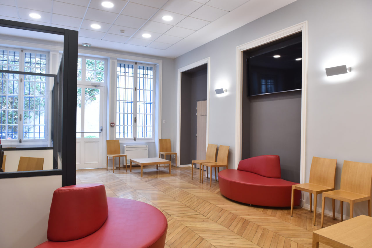
After presenting yourself at the reception desk on level -1, you will be directed to the waiting room on the same level or on level -2 depending on the physician who will perform the examination.
A secretary or operator will direct you to a booth to undress and the physician will ask you to remove any clothing that may interfere with the examination.
The examination takes place lying down or sitting, depending on the organs to be examined. The physician passes a probe over the part of the body to be examined, pressing down to view the internal structures. You may be asked to change your position or breathing in order to get a better view of the organs.
To increase the precision of certain examinations and get as close as possible to the organs to be examined, a special probe protected by a sterile pouch can be inserted into the anus (endorectal ultrasound) to study the prostate, or into the vagina (endovaginal ultrasound) to examine a pregnancy, the uterus, or the ovaries.
The duration of the examination varies depending on the areas to be studied and the level of visibility may vary from one patient to another.
Any preparation you may be required to complete is to enhance the visibility of the organs.
During the examination the physician will take several photographs and the selected images will be printed and attached to the report. They are for illustrative purposes only, as the quality of the reproduction does not in any way allow for a diagnosis to be re-evaluated.
How to retrieve your ultrasound exam.
Your results will be available immediately at the reception desk of the level where you underwent the examination. You will need to wait several minutes to retrieve your examination report. We advise you to ask the secretary how long you will need to wait. If the wait is too long or if you are in a hurry, the report can be sent to you. A copy of the report can also be sent to your physician upon request.

Making an appointment
Certain ultrasound examinations require precautions or preparation:
- Abdominal ultrasound: fast for 3 hours before your appointment. However, don’t forget to take your medication as usual.
- Bladder or pelvic ultrasound: when asked to present with a full bladder, do not urinate for 3 hours before the exam. If you have urinated, drink 4 glasses of water 1 hour beforehand.
Some references on ultrasound by the healthcare team members
- Ultrasound evaluation of the hands and feet in rheumatoid arthritis.
H Guerini, X Ayral, R Campagna, A Feydy, E Pluot, J Rousseau, L Gossec, A Chevrot, M Dougados, JL Drapé. J Radiol. 2010 Jan;91(1 Pt 2):99-110. - Harmonic sonography of rotator cuff tendons: are cleavage tears visible at last?
H Guerini, A Feydy, R Campagna, F Thèvenin, M Fermand, E Pessis, A Chevrot, JL Drapé.
J Radiol. 2008 Mar;89(3 Pt 1):333-8 - Ultrasound of the ankle.
G Morvan, J Busson, M Wybier, P Mathieu. Eur J Ultrasound. 2001 Oct;14(1):73-82. - Ultrasonography of tendons and ligaments of foot and ankle.
G Morvan, P Mathieu, J Busson, M Wybier. J Radiol. 2000 Mar;81(3 Suppl):361-80. - – Osteoarticular ultrasonography of the knee and the hip. M Mathieu; M Wybier; G Morvan
Journal of Radiology (Paris) 2000, 81, 353 – 360 - Sonographic appearance of trigger fingers.
H Guerini, E Pessis, N Theumann, JS Le Quintrec, R Campagna, A Chevrot, A Feydy, JL Drapé. J Ultrasound Med. 2008 Oct;27(10):1407-
